Home
/ Shoulder Muscles Diagram Posterior - Your Complete Guide To Exercises for Shoulder Pain ... / All of these muscles are visible in the diagram pictured.
Shoulder Muscles Diagram Posterior - Your Complete Guide To Exercises for Shoulder Pain ... / All of these muscles are visible in the diagram pictured.
Shoulder Muscles Diagram Posterior - Your Complete Guide To Exercises for Shoulder Pain ... / All of these muscles are visible in the diagram pictured.. The anterior, lateral and posterior deltoid heads. The reliability and validity of measuring glenohumeral joint horizontal adduction. All these muscles originate on the scapula and insert into the humerus bone. The rotator cuff performs multiple functions during shoulder exercises, including glenohumeral abduction, external rotation (er) and internal rotation (ir). Want to learn more about it?
Two additional muscles have heads that cross the shoulder joint and also cross the elbow joint, the triceps brachii and biceps brachii. Anatomy by dr ashwani kumar. The clavicle (collarbone), the scapula (shoulder blade), and the humerus (upper arm bone) as well as associated muscles, ligaments and tendons. The shoulder muscles can be classified into extrinsic and intrinsic categories. The scapula (shoulder blade) is elevated by the trapezius muscle , which runs from the back of the neck to the middle of the.
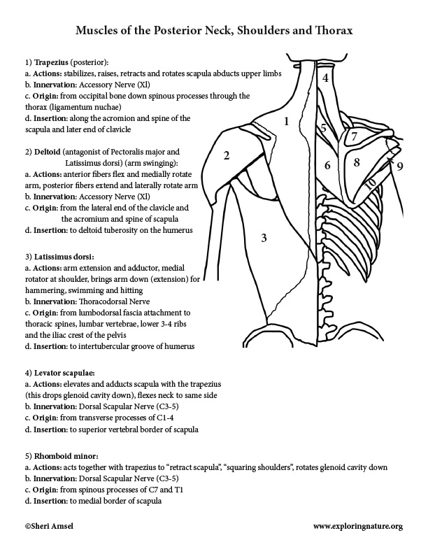
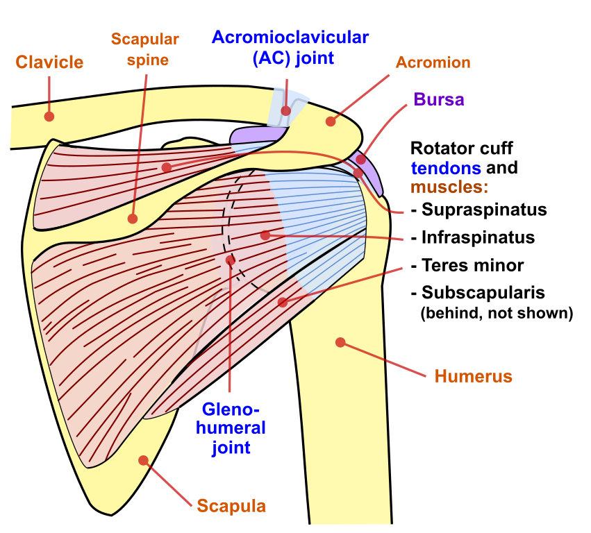
Muscles of the Neck, Shoulders, Chest and Thorax ... from www.exploringnature.org Shoulder muscle anatomy neck muscle anatomy shoulder blade muscles head muscles muscles of the neck anatomy organs anatomy and physiology yoga anatomy human anatomy. The rotator cuff also stabilizes the glenohumeral joint and controls humeral head translations. When only specific fibers are active, can act as a syergist in. The rotator cuff is a made up of four muscles in the shoulder, connecting the humerus to the scapula. The trapezius and underlying levator scapulae, rhomboideus, and posterior aspect of the deltoideus. Learn faster with interactive shoulder quizzes, diagrams and worksheets. The human shoulder is made up of three bones: Muscle strength edit source.
Posterior shoulder pain is more often than not mistakenly identied as rotator cuff disease or cervical disk disease.
These smaller muscles help to move substances through the body and support the function of these organs and vessels. Tutorials on the shoulder muscles (e.g rotator cuff muscles: Nine muscles cross the shoulder joint. Posterior muscles of the arm and forearm. Want to learn more about it? Posterior band of the ighl. Patients with muscle tenderness are diagnosed with myofascial pain. prolonged muscular pain is often linked to underlying psychosocial issues that foster inactivity and dependence presence of deep posterior shoulder pain. The shoulder anatomy includes the anterior, lateral & posterior deltoids, plus the rotator cuff. The rotator cuff also stabilizes the glenohumeral joint and controls humeral head translations. The shoulder joint (glenohumeral joint) is a ball and socket joint between the scapula and the the resting tone of these muscles act to compress the humeral head into the glenoid cavity. The trapezius muscles are the most superficial muscles of the posterior neck and upper trunk; This flow diagram provides an aid to diagnosis of shoulder conditions Posterior shoulder muscle diagram home wiring diagrams.
Acting as a whole, prime mover of arm abduction; Posterior muscles of the arm and forearm. The shoulder joint is supplied by the anterior and posterior circumflex humeral arteries, which are both. Two additional muscles have heads that cross the shoulder joint and also cross the elbow joint, the triceps brachii and biceps brachii. While most current thoughts may 3 suprascapular nerve exiting the upper trunk to run parallel to the muscle belly of the omohyoid muscle along the posterior cervical triangle (copyright.
File:Shoulder joint back-en.svg - Wikimedia Commons from upload.wikimedia.org Infraspinatus and teres minor tendon. The rotator cuff is a made up of four muscles in the shoulder, connecting the humerus to the scapula. Tutorials on the shoulder muscles (e.g rotator cuff muscles: Posterior muscles in the body. Posterior part of the deltoid: Their main function is for the most part, the neck muscles, which move the head and shoulder girdle, are small and straplike. The rotator cuff performs multiple functions during shoulder exercises, including glenohumeral abduction, external rotation (er) and internal rotation (ir). Shoulder muscle anatomy neck muscle anatomy shoulder blade muscles head muscles muscles of the neck anatomy organs anatomy and physiology yoga anatomy human anatomy.
The trapezius muscles are the most superficial muscles of the posterior neck and upper trunk;
When only specific fibers are active, can act as a syergist in. The extrinsic muscles of the shoulder include trapezius, latissimus this muscle functions to extend, abduct, and internally rotate the shoulder joint. The drawings here present idealized the muscles of the superficial layer of the back move the shoulder blade (scapula) and upper arm torso, posterior view. Anterior graphic of the shoulder. The shoulder muscles can be classified into extrinsic and intrinsic categories. Posterior shoulder muscle diagram home wiring diagrams. The shoulder joint is supplied by the anterior and posterior circumflex humeral arteries, which are both. Fleshy triangular muscle forming shoulder muscle mass; Infraspinatus and teres minor tendon. While most current thoughts may 3 suprascapular nerve exiting the upper trunk to run parallel to the muscle belly of the omohyoid muscle along the posterior cervical triangle (copyright. Posterior muscles of the arm and forearm. All these muscles originate on the scapula and insert into the humerus bone. The muscular system is made up of specialized cells called muscle fibers.
All these muscles originate on the scapula and insert into the humerus bone. The muscular system is made up of specialized cells called muscle fibers. Muscles of the shoulder can be divided into two strata: The clavicle (collarbone), the scapula (shoulder blade), and the humerus (upper arm bone) as well as associated muscles, ligaments and tendons. When only specific fibers are active, can act as a syergist in.
FIX YOUR SHOULDER PAIN (Posterior Cuff Active Release ... from i.ytimg.com Shoulder muscle anatomy neck muscle anatomy shoulder blade muscles head muscles muscles of the neck anatomy organs anatomy and physiology yoga anatomy human anatomy. The human shoulder is made up of three bones: Anatomy by dr ashwani kumar. Tutorials on the shoulder muscles (e.g rotator cuff muscles: When only specific fibers are active, can act as a syergist in. Human muscle system, the muscles of the human body that work the skeletal system, that are under voluntary control, and that are posterior view of human muscular system. Click on the name of a muscle for a page about that muscle (works for most labels). Anterior part of the deltoid:
When only specific fibers are active, can act as a syergist in.
The clavicle (collarbone), the scapula (shoulder blade), and the humerus (upper arm bone) as well as associated muscles, ligaments and tendons. Related posts of shoulder muscles labelled diagram. They are also categorized figure 1: The latissimus dorsi also transversely extends and flexes the. Pain in the shoulder joint. Fleshy triangular muscle forming shoulder muscle mass; In order to achieve the maximum release, the patient should lay face up with a lacrosse ball under them. Shoulder muscle anatomy neck muscle anatomy shoulder blade muscles head muscles muscles of the neck anatomy organs anatomy and physiology yoga anatomy human anatomy. The posterior muscles of the shoulder: Muscle strength edit source. Want to learn more about it? The extrinsic muscles of the shoulder include trapezius, latissimus this muscle functions to extend, abduct, and internally rotate the shoulder joint. Nine muscles cross the shoulder joint.
Posterior part of the deltoid: shoulder muscles diagram. The latissimus dorsi also transversely extends and flexes the.
